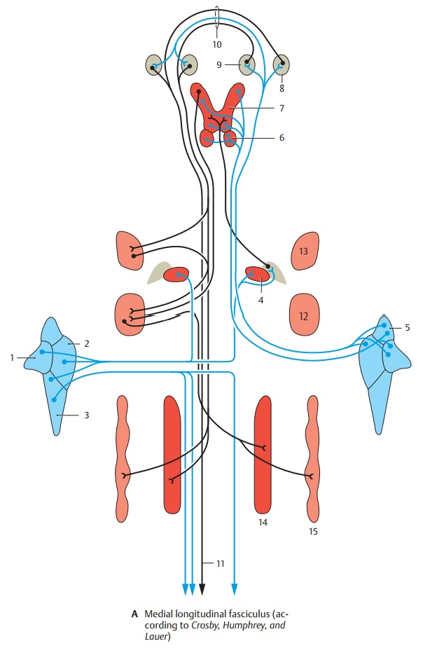
The dorsal portion of the mlf including the mauthner axon continues into the spinal chord while the ventral.Although the medial longitudinal fasciculus was identified in the 1870s, internuclear ophthalmoplegia was first described in 1903.
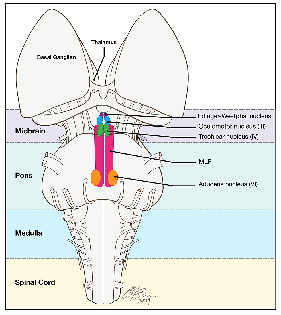
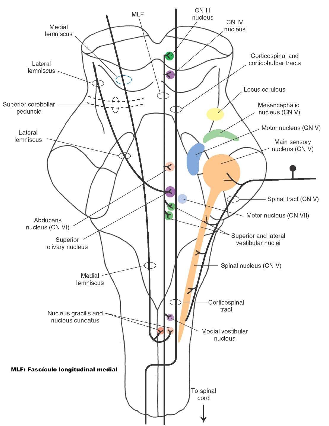
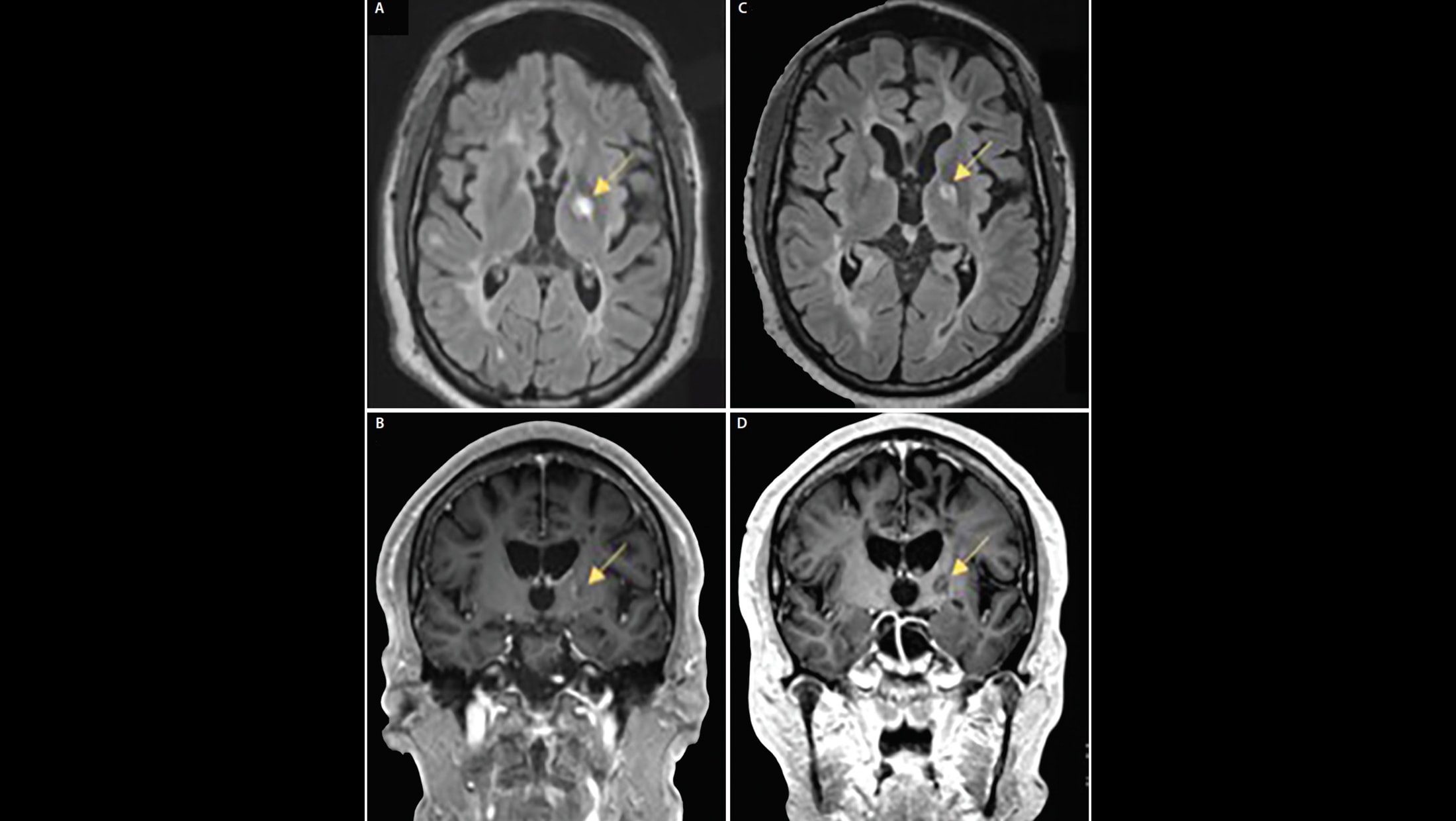
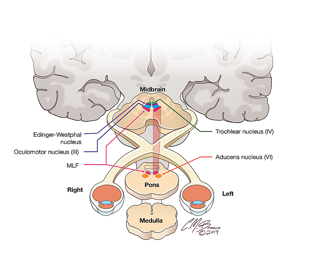
The medial longitudinal fasciculus is located in the dorsomedial aspect of the midbrain tegmentum, adjacent to the ventromedial border of the periventricular gray matter.[4] it connects with the superior colliculus, the vestibular nuclei.The mlf connects the nuclei that control extraocular muscles (abducens, trochlear, and oculomotor nuclei) with one another and with the vestibular nuclei.
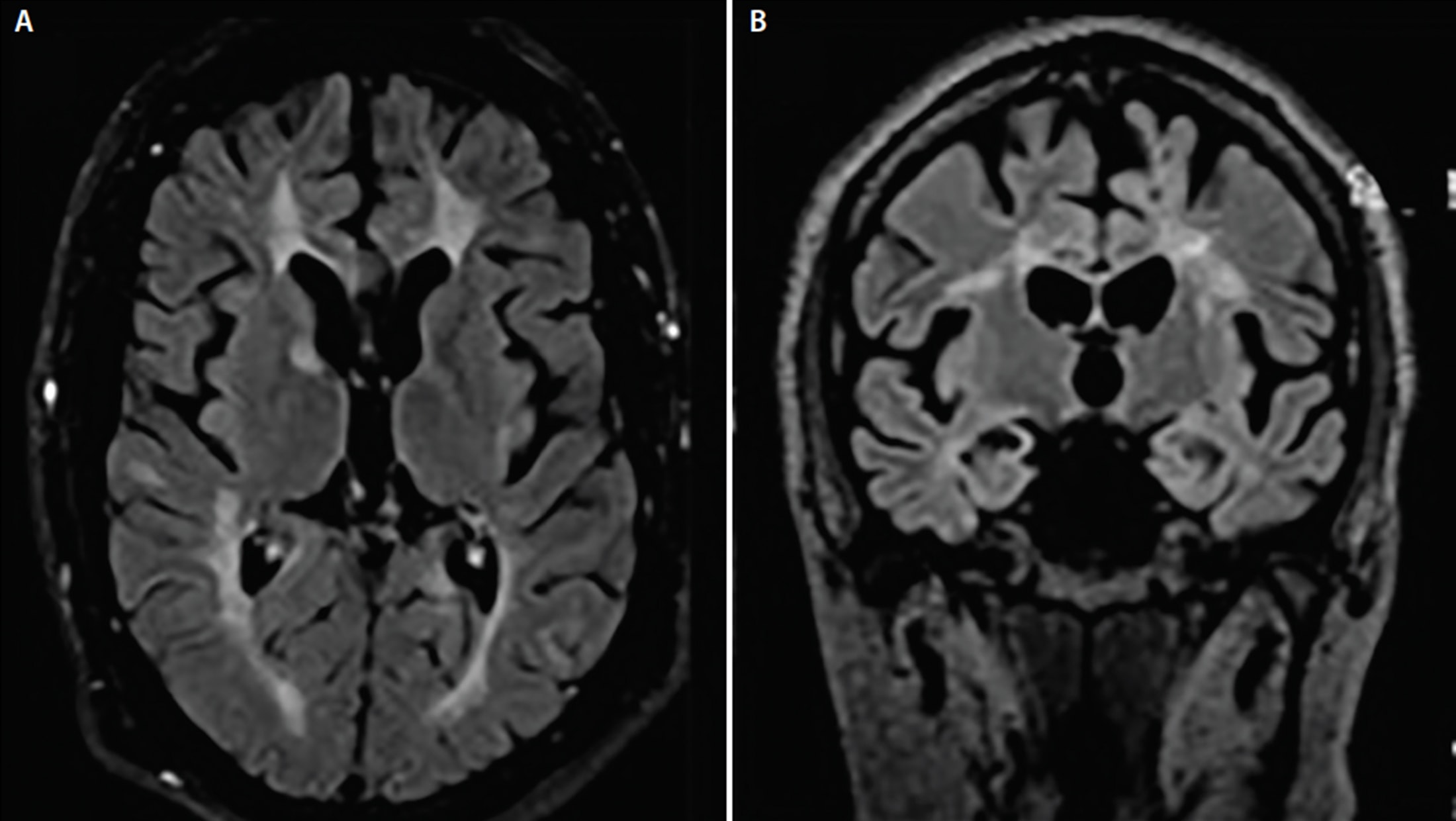
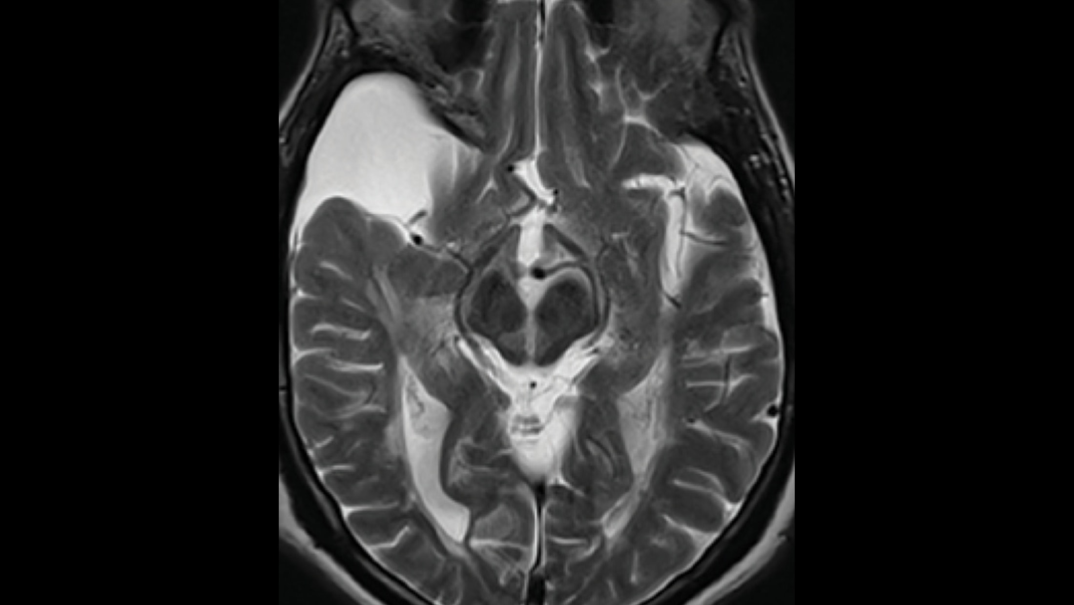
The medial longitudinal fasciculus (mlf) is a highly specialized and organized fiber bundle composed of ascending and descending white matter tracts.The right temporal lobe subarachnoid cyst is an incidental finding.
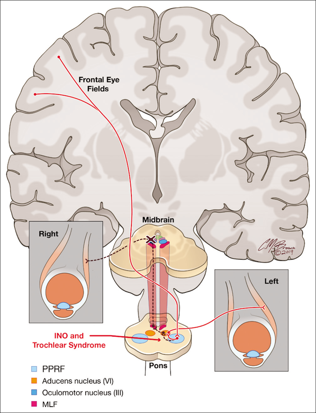
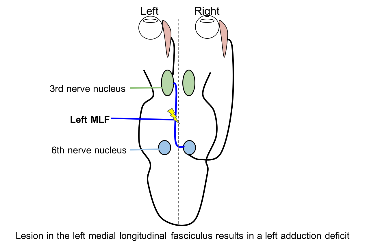
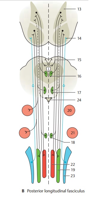
The medial longitudinal fasciculus is a key structure for conjugate horizontal eye movements by relaying signals from the abducens internuclear neurons to the medial rectus subdivision of the contralateral oculomotor nucleus.The medial longitudinal fasciculus (mlf) is pair of longitudinal bundles of white matter that provide a pathway from brain stem to the cervical spinal cord [1,2,3,4].the mlf interacts with eye movement control circuits, which are ascending pathway fibers, and some descending pathway fibers within the brainstem tegmentum involved in the adjustment of horizontal, vertical, and.Medial medullary infarctions (mmis) account for less than 0.5% of all cerebral infarcts and are mostly unilateral.
Learn more or try it out now.
Last update images today Medial Longitudinal Fasciculus
 Ferguson Earns Euro Tour Win, Spot In The Open
Plan ahead in fantasy baseball with help from our forecaster projections. Each day, we will provide an updated preview of the next 10 days for every team, projecting the matchup quality for hitters (overall and by handedness) as well as for base stealers.
This page will be updated daily throughout the season, so be sure to check back often for the latest 10-day outlook.
For our 10-day projections for each team's pitching matchups, click here.